

")
Description
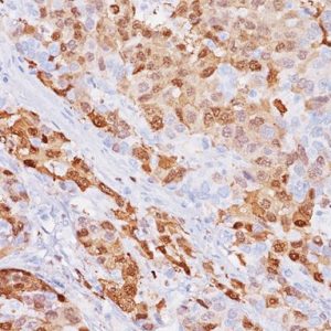
Thyroid transcription factor-1 (TTF-1) is a 38 kDa member of the NKX2 family of homeodomain transcription factors. TTF-1 is mostly detected in primary lung adenocarcinomas and small cell carcinomas (1). TTF-1 can be very useful in lung cancers when used in a panel with Desmoglein 3, p40 and Napsin A antibodies (2-3).
Commercially available thyroid transcription factor-1 (TTF-1) monoclonal antibodies 8G7G3/1 and SPT24 have been shown to have different sensitivities in lung adenocarcinomas (LADC) and lung squamous cell carcinomas (SqCC) (4-6). A study by Masai, et al. demonstrated that SPT24 was much more sensitive than 8G7G3/1 in LADC (72.4% and 65.4% respectively). However, the study demonstrated that SPT24 stained a higher percentage of lung SqCC (16.8% vs. 1%). Higher sensitivity of SPT24 in lung SqCC has also been shown to be heavily influenced by different detection systems (4-5).
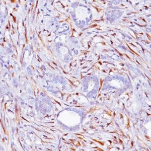
Higher sensitivity for LADC versus lung SqCC can be achieved with SPT24, compared to 8G7G3/1, while retaining specificity, by the use of a cut-off value and optimal antibody titer. In an in-house study, SPT24 was titered to achieve negative staining in normal liver (no cytoplasmic staining observed). A cut-off value of ≥10% of tumor cells positive for TTF-1 with a staining intensity of ≥ 1+ was used to identify TTF-1 positive cases. Using this approach, SPT24 was highly sensitive for LADC (53/60, 88%), compared to 8G7G3/1 (38/60, 63%), with equivalent specificity for both clones versus lung SqCC (2/137, 1.5%).
Use of lung SqCC specific markers, such as Desmoglein 3 and p40, may identify TTF-1 positive cases of squamous cell origin. Additionally, the use of Napsin A may confirm lung adenocarcinoma as the co-expression of Napsin A and TTF-1 in lung cancers has been shown to be more pulmonary specific than either one used alone (7). Finally, unlike clone 8G7G3/1, no cytoplasmic staining in lung cancers has been observed with clone SPT24 (8).
SPECIFICATIONS
Specifications
| INTENDED USE | IVD |
|---|---|
| FORMAT | Concentrate, ONCORE Pro, Predilute, VALENT |
| VOLUME | 0.1 ml, 1.0 ml, 20 ml, 6.0 ml, 60 Tests |
| SOURCE | Mouse Monoclonal |
| CLONE | SPT24 |
| ISOTYPE | IgG1/kappa |
| ANTIGEN | TTF-1 |
| LOCALIZATION | Nuclear |
| POSITIVE CONTROL | Lung adenocarcinoma |
DATASHEETS & SDS
ONCORE PROTOCOL
| ANTIBODY | DOWNLOAD |
|---|---|
| Download Oncore Protocol |
REFERENCES
1. Di Loreto C, et al. Immunocytochemical expression of tissue specific transcription factor-1 in lung carcinoma. J Clin Pathol. 1997 Jan; 50(1):30-2.
2. Tacha D, et al. A 6-antibody panel for the classification of lung adenocarcinoma versus squamous cell carcinoma. Appl Immunohistochem Mol Morphol. 2012 May; 20 (3):201-7.
3. Brown AF, et al. Tissue-preserving antibody cocktails to differentiate primary squamous cell carcinoma, adenocarcinoma, and small cell carcinoma of lung. Arch Pathol Lab Med. 2013 Sep; 137(9):1274-81.
4. Masai K, et al. Expression of squamous cell carcinoma markers and adenocarcinoma markers in primary pulmonary neuroendocrine carcinomas. Appl Immunohistochem Mol Morphol. 2013 Jul; 21(4):292-7.
5. Matoso A, et al. Comparison of thyroid transcription factor-1 expression by 2 monoclonal antibodies in pulmonary and nonpulmonary primary tumors. Appl Immunohistochem Mol Morphol. 2010 Mar; 18(2):142-9.
6. Ordóñez NG. Value of thyroid transcription factor-1 immunostaining in tumor diagnosis: a review and update. Appl Immunohistochem Mol Morphol. 2012 Oct; 20 (5):429-44.


Reviews
There are no reviews yet.