

")
Description
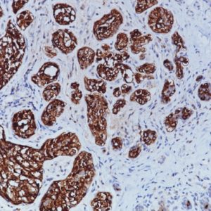
PAX8 antibody is expressed in a high percentage of renal cell carcinomas and ovarian cancers. PAX8 [BC12] has been designed to target restricted epitopes and exhibits higher specificity and provides sharper staining than the PAX8 rabbit polyclonal antibody. PAX8(M) stains nuclei exclusively and does not stain B-cells, nor does it recognize epitopes of pancreatic origin and neuroendocrine cells in stomach and colon. The expression of the mouse monoclonal PAX8 target antigens was found in normal kidney, thyroid and cervix, but was not identified in normal ovary. By western blot, [BC12] has been shown to recognize PAX8 and not PAX2, PAX5 or PAX6 proteins. US Patent 8,852,592 and patents pending.
SPECIFICATIONS
Specifications
| WEIGHT | N/A |
|---|---|
| DIMENSIONS | N/A |
| INTENDED USE | IVD |
| SPECIES REACTIVITY | Cat, Dog, Human, Mouse, Rat |
| SOURCE | Mouse Monoclonal |
| CLONE | BC12, Biocare Clone |
| ISOTYPE | IgG1 |
| ANTIGEN | PAX8 |
| LOCALIZATION | Nuclear |
| POSITIVE CONTROL | Normal kidney, renal cell or serous ovarian carcinomas |
DATASHEETS & SDS
| Download Data Sheet |
| Download RUO Data Sheet for International |
| Download SDS Sheet |
Regulatory Notice: Biocare’s IVD-labeled products comply with US-FDA and European IVDD regulation. Other regions may have additional requirements for such labeling, please contact your local distributor.
REFERENCES
1. Tacha D, Qi W, Zhou D, Bremer R, Cheng L. PAX8 Mouse Monoclonal Antibody [BC12] Recognizes a Restricted Epitope and Is Highly Sensitive in Renal Cell and Ovarian Cancers But Does Not Cross-react With B Cells and Tumors of Pancreatic Origin. Appl Immunohistochem Mol Morphol. Published Ahead-of-Print, May 16, 2012.
2. Tacha D, Zhou D, Cheng L. Expression of PAX8 in Normal and Neoplastic Tissues: A Comprehensive Immunohistochemical Study. Appl Immunohistochem Mol Morphol. 2011 Jul;19(4):293-9.
3. Lotan TL, Ye H, Melamed J, Wu XR, Shih IM, Epstein JI. Immunohistochemical panel to identify the primary site of invasive micropapillary carcinoma. Am J Surg Pathol. 2009 Jul; 33(7):1037-41.
4. Viktorová T, Babjuk M, Dusková J, Stolz J, Goetz P, Mares J. Expression of PAX2 and PAX8 genes in conventional type of renal carcinoma and their role in the tumor prognosis. Diagn Cytopathol. 2008 Aug; 36(8):568-73.
5. Narlis M, Grote D, Gaitan Y, Boualia SK, Bouchard M. Pax2 and Pax8 regulate branching morphogenesis and nephron differentiation in the developing kidney. J Am Soc Nephrol. 2007 Apr; 18(4):1121-9.
6. Moretti L, Medeiros LJ, Kunkalla K et. al. N-terminal PAX8 polyclonal antibody shows cross-reactivity with N-terminal region of PAX5 and is responsible for reports of PAX8 positivity in malignant lymphomas. Mod Pathol 2012 Feb;25(2):231-6.
7. Lorenzo PI, Jimenez Moreno CM, Delgado I et. al. . Immunohistochemical assessment of Pax8 expression during pancreatic islet development and in human neuroendocrine tumors. Histochem Cell Biol 2011 Nov;136(5):595-607.
8. Center for Disease Control Manual. Guide: Safety Management, NO. CDC-22, Atlanta, GA. April 30, 1976 “Decontamination of Laboratory Sink Drains to Remove Azide Salts.”
9. National Committee for Clinical Laboratory Standards (NCCLS). Protection of laboratory workers from infectious diseases transmitted by blood and tissue; proposed guideline. Villanova, PA 1991; 7(9).


Reviews
There are no reviews yet.