

Description
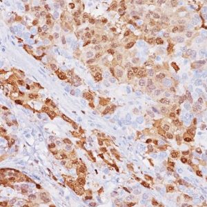
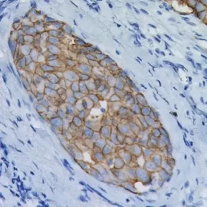
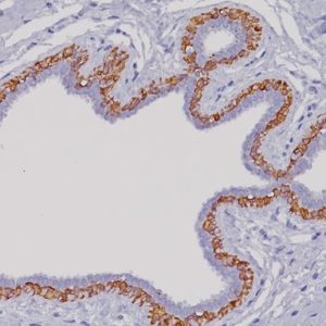
The emergence of new strains of resistant mycobacterium tuberculosis has created interest in clinical diagnosis. Studies have shown immunohistochemical and immunofluorescent techniques to be superior to conventional special stains in the detection of mycobacterium. Demonstrating mycobacterial antigens is useful in establishing mycobacterial etiology and can be used as an alternative method to the conventional Ziehl-Neelsen method. Studies have shown that Mycobacterium tuberculosis antibody is reactive with other mycobacteria species, but is not reactive with E. coli K12, Salmonella typhimurium, Pseudomonas aeruginosa, Streptococcus (group B), Candida albicans and Neisseria meningitides.
SPECIFICATIONS
Specifications
| WEIGHT | N/A |
|---|---|
| DIMENSIONS | N/A |
| INTENDED USE | RUO |
| SPECIES REACTIVITY | Human |
| SOURCE | Rabbit Polyclonal |
| CLONE | N/A |
| ISOTYPE | N/A |
| ANTIGEN | Mycobacterium tuberculosis |
| LOCALIZATION | N/A |
| POSITIVE CONTROL | Mycobacterium tuberculosis infected tissue |
DATASHEETS & SDS
OUS ONLY IVD
REFERENCES
1. Radhakrishnan VV, et al. Immunohistochemical demonstration of mycobacterial
antigens in intracranial tuberculoma. Indian J Exp Biol. 1991 Jul;29(7):641-4.
2. Center for Disease Control Manual. Guide: Safety Management, NO. CDC-22,
Atlanta, GA. April 30, 1976 “Decontamination of Laboratory Sink Drains to Remove
Azide Salts.”
3. Clinical and Laboratory Standards Institute (CLSI). Protection of Laboratory
Workers from Occupationally Acquired Infections; Approved guideline-Third Edition
CLSI document M29-A3 Wayne, PA 2005.


Reviews
There are no reviews yet.