

Description
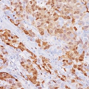
MART-1 recognizes a protein of 18 kDa, identified at MART-1 (Melanoma Antigen Recognized by T cells) (1). MART-1 is a useful addition to melanoma panels as it is apparently specific for melanocytic lesions (1,2). Studies have shown that MART-1 is more sensitive than HMB45 when labeling metastatic melanomas (3). This MART-1 cocktail does not stain steroid tumors like Melan A [103] does. Tyrosinase has been shown a more sensitive marker in recognizing melanoma when compared to HMB45 and MART-1. It has also been shown to label a higher percentage of desmoplastic melanomas than HMB45 (1). The combination of MART-1 and Tyrosinase aids in identifying metastatic melanoma in sentinel lymph nodes (4).
Microscopic evaluation of mitotic figures on H&E is a routine procedure in the assessment of the tumor grades (5). However, the counting of mitosis is manual and time consuming with assorted difficulties as well as variabilities between interobserver assessments (6). Histone H3 phosphorylation at Serine10 (pHH3) is in association with mitotic chromatin condensation in late G2 and M phase of the cell cycle. pHH3 can distinguish mitosis from apoptotic nuclei (7). The immunohistochemical staining of Serine10-pHH3 has been reported to be comparable to mitotic figures in the H&E section (8-11). The combination of monoclonal anti-pHH3 with MART-1 and Tyrosinase would offer an advantage of specific epitopes for melanoma diagnosis and mitosis counting.
SPECIFICATIONS
Specifications
| INTENDED USE | IVD |
|---|---|
| FORMAT | Predilute |
| VOLUME | 6.0 ml |
| ANTIGEN | MART-1, PhosphoSer10 of Histone H3, Tyrosinase |
| ISOTYPE | IgG, IgG2a, IgG2b |
| SOURCE | Mouse Monoclonal, Rabbit Monoclonal |
| CLONE | BC37, Biocare Clone, M2-7C10 + M2-9E3, T311 |
| BY LETTER | M |
DATASHEETS & SDS
REFERENCES
1. Orchard G. Evaluation of melanocytic neoplasms: application of a pan-melanoma antibody cocktail. Br J Biomed Sci. 2002;59(4):196- 202.
2. Blessing K, et al. Comparison of immunohistochemical staining of the novel antibody Melan-A with S100 protein and HMB-45 in malignant melanoma and melanoma variants. Histopathology. 1998 Feb; 32 (2):139-46.
3. Cook MG, et al. The development of optimal pathological assessment of sentinel lymph nodes for melanoma. J Pathol. 2003 Jul;200(3):314-9.
4. Miettinen M, et al. Microphthalmia transcription factor in the immunohistochemical diagnosis of metastatic melanoma: comparison with four other melanoma markers. Am J Surg Pathol. 2001 Feb;25(2):205-11.
5. Jannink I, et al. Comparison of the prognostic value of four methods to assess mitotic activity in 186 invasive breast cancer patients: classical and random mitotic activity assessments with correction for volume percentage of epithelium. Hum Pathol. 1995 Oct;26(10):1086-92.
6. Yadav KS, et al. Assessment of interobserver variability in mitotic figure counting in different histological grades of oral squamous cell carcinoma. J Contemp Dent Pract. 2012 May 1;13(3):339-44.
7. Ladstein RG, et al. Prognostic importance of the mitotic marker phosphohistone H3 in cutaneous nodular melanoma. J Invest Dermatol. 2012 Apr;132(4):1247-52.
8. Thareja S, et al. Analysis of tumor mitotic rate in thin metastatic melanomas compared with thin melanomas without metastasis using both the hematoxylin and eosin and anti-phosphohistone 3 IHC stain. Am J Dermatopathol. 2014 Jan;36(1):64-7.
9. Ikenberg K, et al. Immunohistochemical dual staining as an adjunct in assessment of mitotic activity in melanoma. J Cutan Pathol. 2012 Mar;39(3):324-30.
10. Casper DJ, et al. Use of anti-phosphohistone H3 immunohistochemistry to determine mitotic rate in thin melanoma. Am J Dermatopathol. 2010 Oct;32(7):650-4.
11. Fulton R, Tacha D. Use of a Novel Rabbit Monoclonal Phospho- Histone H3 (Ser10) versus H&E Mitotic Count in Melanoma. Mod Pathol 2016;29: (Suppl 2) 128A.
12. Center for Disease Control Manual. Guide: Safety Management, NO. CDC-22, Atlanta, GA. April 30, 1976 “Decontamination of Laboratory Sink Drains to Remove Azide Salts.”
13. Clinical and Laboratory Standards Institute (CLSI). Protection of Laboratory Workers from Occupationally Acquired Infections; Approved Guideline-Fourth Edition CLSI document M29-A4 Wayne, PA 2014.


Reviews
There are no reviews yet.