

Description
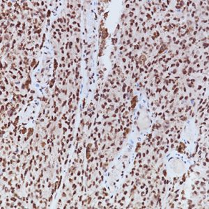
Glial Fibrillary Acidic Protein antibody reacts with human GFAP and does not react with other intermediate filaments. Anti-GFAP stains astrocytes, ependymal cells and corresponding tumors. Studies have shown that GFAP may be useful for distinguishing neoplasms of astrocytic origin. GFAP may also be useful in differentiating gliomas from metastatic lesions in the brain. Neuroblastomas, Schwannomas, as well as extra-CNS tumors are not labeled. Negative staining has been observed with lymphatic tissue, muscle, gastrointestinal tract, liver, kidney, pancreas and bladder. Use of a monoclonal antibody typically will increase specificity and eliminate lot-to-lot variation seen with polyclonals.
SPECIFICATIONS
Specifications
| WEIGHT | N/A |
|---|---|
| DIMENSIONS | N/A |
| INTENDED USE | IVD |
| SPECIES REACTIVITY | Human, Mouse, Rat |
| SOURCE | Mouse Monoclonal |
| CLONE | GA-5 |
| ISOTYPE | IgG1 |
| ANTIGEN | Glial Fibrillary Acidic Protein |
| LOCALIZATION | Cytoplasmic |
| POSITIVE CONTROL | Normal brain or astrocytoma |
DATASHEETS & SDS
REFERENCES
1. Huang MC, Kubo O, Tajika Y, Takakura K. A clinico-immunohistochemical study of giant cell glioblastoma. 1996 Apr;13(1):11-16.
2. Xu KP, Liu SL, Ni C. Immunohistochemical evidence of neuronal and glial differentiation in retinoblastoma. Br J Opthalmol 1995 Aug;79(8):771-776.
3. Center for Disease Control Manual. Guide: Safety Management, NO. CDC-22, Atlanta, GA. April 30, 1976 “Decontamination of Laboratory Sink Drains to Remove Azide Salts.”
4. National Committee for Clinical Laboratory Standards (NCCLS). Protection of laboratory workers from infectious diseases transmitted by blood and tissue; proposed guideline. Villanova, PA 1991;7(9). Order code M29-P.


Reviews
There are no reviews yet.