

Description
FOXP3 [86D] is a mouse monoclonal antibody that is intended for laboratory use in the qualitative identification of FOXP3 protein by immunohistochemistry (IHC) in formalin-fixed paraffin-embedded (FFPE) human tissues. The clinical interpretation of any staining or its absence should be complemented by morphological studies using proper controls and should be evaluated within the context of the patient’s clinical history and other diagnostic tests by a qualified pathologist.
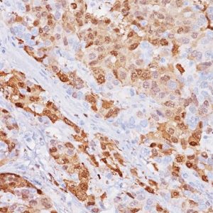
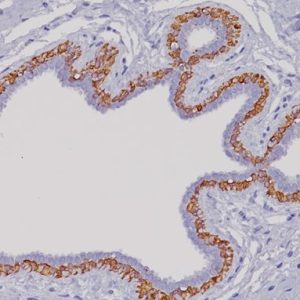
FOXP3 is a forkhead transcription factor family member involved in T-cell regulation, activation and differentiation. FOXP3 has been shown to be a master control gene for the development and function of CD4+/CD25+ regulatory T-cells. In IHC, FOXP3 has been shown to be a specific marker for adult T-cell leukemia/lymphoma (1). In melanoma and in breast and lung cancers, high numbers of circulating regulatory T cells have been associated with disease progression (2-5). Conversely, the infiltration of FOXP3+ regulatory T cells into invasive tumors has also been reported to be associated with survival in a variety of cancers (2-7). In colon cancers, a high frequency of FOXP3+ infiltrates has shown to be a positive indicator (6-7). Patients with high FOXP3 expression in Crohn’s disease have shown a better response to infliximab therapy (8). In allography recipients, FOXP3 cell levels may also be useful in improving post-transplant management (9).
SPECIFICATIONS
Specifications
| BY LETTER | F |
|---|---|
| INTENDED USE | IVD |
| FORMAT | Concentrate, Predilute |
| CLONE | 86D |
| VOLUME | 0.1 ml, 1.0 ml, 6.0 ml |
| ISOTYPE | IgG1 |
| LOCALIZATION | Nuclear |
| POSITIVE CONTROL | Colon cancer and tonsil |
| SOURCE | Mouse Monoclonal |
| SPECIES REACTIVITY | Human, Others Not Tested |
DATASHEETS & SDS
REFERENCES
1. Roncador G, et al. FOXP3, a selective marker for a subset of adult T-cell leukaemia/lymphoma. Leukemia. 2005 Dec; 19(12):2247-53.
2. Gerber AL, et al. High expression of FOXP3 in primary melanoma is associated with tumor progression. Br J Dermatol. 2014 Jan; 170(1):103-9.
3. Ali HR, et al. Association between CD8+ T-cell infiltration and breast cancer survival in 12,439 patients. Ann Oncol. 2014 Aug; 25(8):1536- 43.
4. Liu H, et al. Tumor-infiltrating lymphocytes predict response to chemotherapy in patients with advance non-small cell lung cancer. Cancer Immunol Immunother. 2012 Oct; 61(10):1849-56.
5. Tao H, et al. Density of tumor-infiltrating FOXP3+ T cells as a response marker for induction chemoradiotherapy and a potential prognostic factor in patients treated with trimodality therapy for locally advanced non-small cell lung cancer. Ann Thorac Cardiovasc Surg. 2014; 20(6):980-6.
6. Ling A, et al. The intratumoural subsite and relation of CD8(+) and FOXP3(+) T lymphocytes in colorectal cancer provide important prognostic clues. Br J Cancer. 2014 May 13; 110(10):2551-9.
7. Frey DM, et al. High frequency of tumor-infiltrating FOXP3(+) regulatory T cells predicts improved survival in mismatch repairproficient colorectal cancer patients. Int J Cancer. 2010 Jun 1; 126(11):2635-43.
8. Sloan S, et al. FOXP3+ regulatory T-cell counts correlate with histological response in Crohn’s colitis treated with infliximab. Pathol Int. 2014 Dec; 64(12):624-7.
9. Stenard F, et al. Decreases in circulating CD4+CD25hiFOXP3+ cells and increases in intragraft FOXP3+ cells accompany allograft rejection in pediatric liver allograft recipients. Pediatr Transplant. 2009 Feb; 13(1):70-80.
10. Center for Disease Control Manual. Guide: Safety Management, NO. CDC-22, Atlanta, GA. April 30, 1976 “Decontamination of Laboratory Sink Drains to Remove Azide Salts.”
11. Clinical and Laboratory Standards Institute (CLSI). Protection of Laboratory Workers from Occupationally Acquired Infections; Approved Guideline-Fourth Edition CLSI document M29-A4 Wayne, PA 2014.


Reviews
There are no reviews yet.