

Description
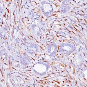
Cytokeratin 5 antibody rabbit is a type II intermediate filament protein that is expressed in active basal layers of most stratified squamous epithelia. CK5 is expressed in many non-keratinizing stratified squamous epithelia as well as basal cells in prostate glands and myoepithelial cells in mammary glands. In a published study, rabbit monoclonal CK5 antibody was compared to mouse monoclonal CK5/6. CK5 was 84% sensitive and 100% specific for lung SqCC when compared to CK5/6 (80% sensitivity and 97% specificity). The CK5 predilute has been optimized for lung squamous cell carcinoma; other tumors have not been tested.
SPECIFICATIONS
Specifications
| WEIGHT | N/A |
|---|---|
| DIMENSIONS | N/A |
| INTENDED USE | IVD |
| SPECIES REACTIVITY | Human |
| SOURCE | Rabbit Monoclonal |
| CLONE | EP42 |
| ISOTYPE | N/A |
| ANTIGEN | C-terminal region of human CK5 |
| LOCALIZATION | Cytoplasmic |
| POSITIVE CONTROL | Lung squamous cell carcinoma, some breast cancers, or normal prostate or skin |
DATASHEETS & SDS
| Download DS Data Sheet |
| Download RUO Data Sheet for International |
| Download SDS Sheet |
Regulatory Notice: Biocare’s IVD-labeled products comply with US-FDA and European IVDD regulation. Other regions may have additional requirements for such labeling, please contact your local distributor.
REFERENCES
1. Mukhopadhyay S, Katzenstein AL. Subclassification of non-small cell lung carcinomas lacking morphologic differentiation on biopsy specimens: Utility of an immunohistochemical panel containing TTF-1, Napsin A, p63, and CK5/6. Am J Surg Pathol. 2011 Jan; 35(1):15-25.
2. Tacha D, Yu C, Haas T. TTF-1, Napsin A, p63, TRIM29, Desmoglein-3 and CK5: An Evaluation of Sensitivity and Specificity and Correlation of Tumor Grade for Lung Squamous Cell Carcinoma vs. Lung Adenocarcinoma. Modern Pathology; USCAP, 2011 (Abstract Accepted)
3. Tacha D, Zhou D, Henshall-Powell RL¹. Distinguishing Adenocarcinoma from Squamous Cell Carcinoma in Lung Using Double Stains p63+ CK5 and TTF-1 + Napsin A. Modern Pathology; Pathology Volume 23, Supplement 1, Feb 2010; Abstract 1852, page 222A.
4. Terry J, et al. Optimal immunohistochemical markers for distinguishing lung adenocarcinomas from squamous cell carcinomas in small tumor samples. Am J Surg Pathol. 2010 Dec; 34(12):1805-11.
5. Kargi A, Gurel D, Tuna B. The diagnostic value of TTF-1, CK 5/6, and p63 immunostaining in classification of lung carcinomas. Appl Immunohistochem Mol Morphol. 2007 Dec; 15(4):415-20.
6. Miettinen M, Sarlomo-Rikala M. Expression of calretinin, thrombomodulin, keratin
5, and mesothelin in lung carcinomas of different types: an immunohistochemical analysis of 596 tumors in comparison with epithelioid mesotheliomas of the pleura. Am J Surg Pathol. 2003 Feb; 27(2):150-8.
7. Bocker W, et al. Common adult stem cells in the human breast give rise to glandular and myoepithelial cell lineages: a new cell biological concept. Lab Invest. 2002 Jun; 82 (6):737-46.
8. Center for Disease Control Manual. Guide: Safety Management, NO. CDC-22, Atlanta, GA. April 30, 1976 “Decontamination of Laboratory Sink Drains to Remove Azide Salts.”
9. Clinical and Laboratory Standards Institute (CLSI). Protection of Laboratory workers from occupationally Acquired Infections; Approved guideline-Third Edition CLSI document M29-A3 Wayne, PA 2005


Reviews
There are no reviews yet.