

Description
IHC markers CK5, CK14, p63, CK7 and CK18 complement morphological evaluation of breast lesions, due to the differential expression of the luminal (CK7/18) vs. basal and myoepithelial markers (CK5/14, p63). Usual ductal hyperplasia is associated with positive basal cells markers, intermixed with positive luminal cells. Most atypical ductal hyperplasia and low grade ductal carcinoma in situ cases are basal marker negative and luminal marker positive. These antibodies, in combination with hematoxylin and eosin (H&E), have been shown to significantly increase diagnostic inter-observer agreement among pathologists.
CK5/14 + p63 + CK7/18 is comprised of mouse monoclonal anti-CK5, anti-CK14, and anti-p63 antibodies and rabbit monoclonal anti-CK7 and anti-CK18 antibodies. CK5 and CK14 are high molecular weight keratins expressed in the cytoplasm of basal cells and myoepithelium of breast tissue (1-4). p63 is a transcription factor present in the nuclei of myoepithelial cells (2,4). In contrast, CK7 and CK18 are low molecular weight cytokeratins primarily expressed in luminal cells of the breast (1-3).
CK5, CK14, p63, CK7 and CK18 have routinely been used as IHC markers to complement morphological evaluation in the assessment of breast lesions, due to the differential expression of the luminal vs. basal and myoepithelial markers (1-5). Cases of usual ductal hyperplasia (UDH) have been associated with expression of the basal cell markers, intermixed with cells expressing the keratins of luminal cells (1-2,6-10). Most cases of atypical ductal hyperplasia (ADH) and low grade ductal carcinoma in situ (LG-DCIS) were negative for the basal markers and exhibited an immunophenotype indicative of luminal cells (1,5-8). Additionally, the basal phenotype has been shown to be characterized by luminal expression of the basal and myoepithelial markers, using a cocktail of CK5, CK14 and p63 (11-13).
IHC, using CK5, CK14, p63, CK7 and CK18 antibodies, evaluated in combination with hematoxylin and eosin (H&E), has been shown to significantly increase inter-observer agreement amongst pathologists, compared to H&E alone (14).
Free White Paper: Classification & Cocktails: Atypical Ductal Hyperplasia
SPECIFICATIONS
Specifications
| INTENDED USE | IVD |
|---|---|
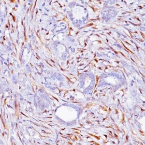
| POSITIVE CONTROL | Breast Carcinoma |
| SPECIES REACTIVITY | Human |
| CLONE | 4A4, BC1, Biocare Clone, EP30, LL002, XM26 |
| SOURCE | Mouse monoclonal, Mouse monoclonal, Mouse monoclonal, Rabbit monoclonal, Rabbit monoclonal |
| ISOTYPE | IgG, IgG1/kappa, IgG2a/kappa, IgG3 |
| ANTIGEN | CK14, CK18, CK5, CK7, p63 |
| LOCALIZATION | Cystoplasmic, Cytoplasmic, Nuclear |
| STAINING | Brown (DAB), Brown (DAB), Brown (DAB), Red (Warp Red), Red (Warp Red) |
ONCORE PROTOCOL
| ANTIBODY | DOWNLOAD |
|---|---|
| Download Oncore Protocol |
REFERENCES
1. Hicks DG. Appl Immunohistochem Mol Morph. 2011 Dec; 19(6):501-05.
2. Jain RK, et al. Mod Pathol. 2011 Jul; 24(7):917-23.
3. Tacha DE, et al. Mod Pathol. 2009 Jan; 22(Suppl 1s):388A.
4. Moriya T, et al. Med Mol Morphol. 2006 Mar; 39(1):8-13.


Reviews
There are no reviews yet.