

Description
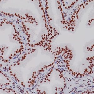
CD4 is expressed in a T-cell subset (helper/inducer) and is found in approximately 80% of thymocytes and in 45% of peripheral blood lymphocytes. CD4 is expressed in the majority of T-cell lymphomas including mycosis fungoides, a common form of cutaneous T-cell lymphoma (1).
CD8 has been shown to be an important marker in the analysis of T-cell mediated inflammatory dermatoses and is also useful for analysis of mycosis fungoides (2-4).
CD8 can be used in panels with CD4, CD56, TIA-1 to aid in identifying subsets of inflammatory skin diseases (4). CD4 and CD8 have also been shown to be valuable in squamous cell cervical cancer and gastric mucosa in HIV infection (5-7). The combination of CD4(+) and CD8(-) is helpful in distinguishing mycosis fungoides and can be used in a panel of CD2(+), CD3(+) and CD7(-/+) (1-3). Multiplex IHC may also give distinct advantages if ratios and/or cell counts on a single slide are desired.
SPECIFICATIONS
Specifications
| INTENDED USE | IVD |
|---|---|
| FORMAT | Predilute |
| VOLUME | 6.0 ml |
| SOURCE | Mouse Monoclonal, Rabbit Monoclonal |
| CLONE | 4B12, SP16 |
| ISOTYPE | IgG, IgG1/kappa |
| ANTIGEN | CD4, CD8 |
| LOCALIZATION | Cell surface |
| POSITIVE CONTROL | Mycosis fungoides and normal tonsil |
| SPECIES REACTIVITY | Human; others not tested |
DATASHEETS & SDS
REFERENCES
1. Boone SL, Guitart J, Gerami P. Follicular mycosis fungoides: a histopathologic, immunohistochemical, and genotypic review. G Ital Dermatol Nenereol. 2008 Dec;143 (6):409-14.
2. Hodak E, et al. CD4/CD8 double-negative epidermotropic cutaneous T-cell lymphoma: an immunohistochemical variant of mycosis fungoides. J Am Acad Dermatol. 2006 Aug;55(2):276-84.
3. Tirumalae R, Panjwani PK. Origin Use of CD4, CD8, and CD1a Immunostains in Distinguishing Mycosis Fungoides from its Inflammatory Mimics: A Pilot Study. Indian J Dermatol. 2012 Nov;57(6):424-7.
4. Harvell JD, Nowfar-Rad M, Sundram U. An immunohistochemical study of CD4,
CD8, TIA-1 and CD56 subsets in inflammatory skin disease. J Cutan Pathol. 2003 Feb;30(2):108-13.
5. Shi Z, et al. Frequency, distribution of CD4+, CD8+ T cells and expression of CD38 in gastric mucosa of HIV infections. Za Zhi. 2009 Aug;23(4):261-4.
6. Shah W, et al. A reversed CD4/CD8 ratio of tumor-infiltrating lymphocytes and a high percentage of CD4(+)FOXP3(+) regulatory T cells are significantly associated with clinical outcome in squamous cell carcinoma of the cervix. Cell Mol Immunol. 2011 Jan;8(1):59-66.
7. Barth TF, et al. Primary gastric apoptosis-rich T-cell lymphoma co-expressing CD4, CD8, and cytotoxic molecules. Virchows Arch. 2000 Apr; 436(4):357-64.
8. Williamson SL, et al. New monoclonal antibodies to the T cell antigens CD4 and CD8. Production and characterization in formalin-fixed paraffin-embedded tissue. Am J Pathol. 1998 Jun; 152(6):1421-6.
9. Center for Disease Control Manual. Guide: Safety Management, NO. CDC-22, Atlanta, GA. April 30, 1976 “Decontamination of Laboratory Sink Drains to Remove Azide Salts.”
10. Clinical and Laboratory Standards Institute (CLSI). Protection of Laboratory Workers from Occupationally Acquired Infections; Approved Guideline-Fourth Edition CLSI document M29-A4 Wayne, PA 2014.


Reviews
There are no reviews yet.