

Description
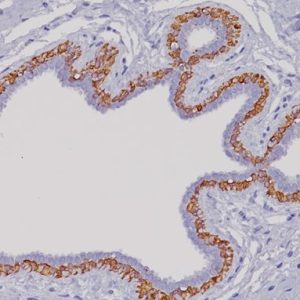
CD1a is a protein of 43 to 49 kDa and is expressed on dendritic cells and cortical thymocytes (1,2). CD1a [O10] staining has been shown to be useful in the differentiation of Langerhans cells from interdigitating cells. It has also proved useful for phenotyping Langerhans cell histiocytosis (2,3). CD1a may be a novel biomarker for Barrett’s metaplasia, and its expression may help to predict the prognosis of this pathology (4).
SPECIFICATIONS
Specifications
| INTENDED USE | IVD |
|---|---|
| FORMAT | Concentrate, Predilute, VALENT |
| VOLUME | 0.1 ml, 0.5 ml, 20 ml, 6.0 ml |
| SOURCE | Mouse Monoclonal |
| CLONE | O10 |
| ISOTYPE | IgG1/kappa |
| ANTIGEN | CD1a |
| LOCALIZATION | Cell membrane and cytoplasm |
| POSITIVE CONTROL | Skin |
| SPECIES REACTIVITY | Human; others not tested |
DATASHEETS & SDS
REFERENCES
1. Krenacs L, et al. Immunohistochemical detection of CD1A antigen in formalin-fixed and paraffin-embedded tissue sections with monoclonal O10. J Pathol. 1993 Oct;171
(2):99-104.
2. Fivenson DP, et al. Distinctive dendritic cell subsets expressing factor XIIIa, CD1a, CD1b and CD1c in mycosis fungoides and psoriasis. J Cutan Pathol. 1995 Jun;22
(3):223-8.
3. Emile JF, et al. Langerhans’ cell histiocytosis. Definitive diagnosis with the use of monoclonal antibody O10 on routinely paraffin-embedded samples. Am J Surg Pathol.
1995 Jun;19(6):636-41.
4. Cappello F, et al. CD1a expression by Barrett’s metaplasia of gastric type may help to predict its evolution towards cancer. Br J Cancer. 2005 Mar 14;92(5):888-90.
5. Center for Disease Control Manual. Guide: Safety Management, NO. CDC-22, Atlanta, GA. April 30, 1976 “Decontamination of Laboratory Sink Drains to Remove Azide Salts.”
6. Clinical and Laboratory Standards Institute (CLSI). Protection of Laboratory Workers from Occupationally Acquired Infections; Approved Guideline-Fourth Edition CLSI document M29-A4 Wayne, PA 2014.


Reviews
There are no reviews yet.