

Description
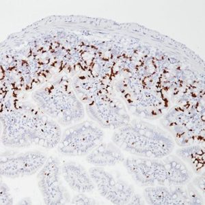
CD138 / syndecan-1 protein backbone is a single chain molecule of 30.5 kDa. Five putative GAG attachment sites exist in the extracellular domain. GAG fine structure appears to reflect the cellular source of the syndecan. Expression of CD138 antibody in human hematopoietic cells is restricted to plasma cells in normal bone marrow. Early B-cell precursors in human bone marrow are CD138 negative. CD138 may aid in distinguishing between viable myeloma cells vs. apoptotic cells. CD138 is also expressed in endothelial cells, fibroblasts, keratinocytes and normal hepatocytes.
Antigen detection in tissues and cells is a multi-step immunohistochemical process. The initial step binds the primary antibody to its specific epitope. After labeling the antigen with a primary antibody, a secondary antibody is added to bind to the primary antibody. An enzyme label is then added to bind to the secondary antibody; this detection of the bound antibody is evidenced by a colorimetric reaction.
SPECIFICATIONS
Specifications
| WEIGHT | N/A |
|---|---|
| DIMENSIONS | N/A |
| INTENDED USE | IVD |
| SPECIES REACTIVITY | Human |
| SOURCE | Mouse Monoclonal |
| CLONE | B-A38 |
| ISOTYPE | IgG1 |
| ANTIGEN | CD138 (Syndecan-1) |
| LOCALIZATION | Cell Membrane |
| POSITIVE CONTROL | Tonsil |
DATASHEETS & SDS
| Download Data Sheet |
| Download RUO Data Sheet |
| Download SDS Sheet |
Regulatory Notice: Biocare’s IVD-labeled products comply with US-FDA and European IVDD regulation. Other regions may have additional requirements for such labeling, please contact your local distributor.
REFERENCES
1. Sun RX, et al. J Immunol Methods. 1997 Jun; 205(1):73-9.
2. Carbone A, et al. Blood. 1997 May; 89(10):3787-94.
3. Jourdan M, et al. Br J Haematol. 1998 Mar; 100(4):637-46.
4. Sebestyén A, et al. Br J Haematol. 1996 Feb; 104(2):412-9.
5. Inki F, Jalkanen M. Ann Med. 1996 Feb; 28(1):63-7.


Reviews
There are no reviews yet.