

Description
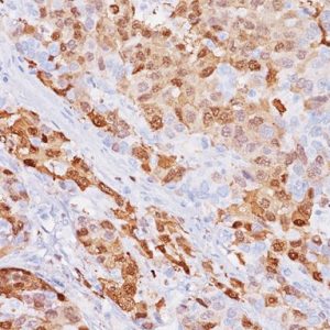
Amyloidosis is a heterogeneous group of disorders characterized by extracellular deposition of abnormal protein fibrils, which are derived from different proteins. Amyloid P antibody reacts with amyloid deposits in all tissues including kidney, rectum and brain. The application of Congo Red, Amyloid P antibody and Amyloid A antibody in tissues with amyloid deposits has been shown to be superior to Congo Red and other histochemical stains. Small and minute amounts of amyloid can be detected with both Amyloid P and Amyloid A antibodies and thus could aid in allowing earlier treatment before organ damage has occurred.
ADDITIONAL INFORMATION
Additional Information
| WEIGHT | N/A |
|---|---|
| DIMENSIONS | N/A |
| INTENDED USE | IVD |
| SOURCE | Rabbit Polyclonal |
| SPECIES REACTIVITY | Human |
| CLONE | N/A |
| ISOTYPE | N/A |
| POSITIVE CONTROL | Amyloid deposits in kidney, or other amyloid-infiltrated tissue |
| ANTIGEN | Amyloid P |
DATASHEETS & SDS
| Download Data Sheet |
| Download RUO Data Sheet |
| Download SDS Sheet |
Regulatory Notice: Biocare’s IVD-labeled products comply with US-FDA and European IVDD regulation. Other regions may have additional requirements for such labeling, please contact your local distributor.
REFERENCES
1. Suwabe H, et al. Pathol Int. 1999 May; 49(5):391-402. 2. Cui D, et al. Pathol Int. 1998 May; 48(5):362-7. 3. Wagrowska-Danilewicz M, Danilewicz M. Acta Histochem. 1996 Jul; 98(3):301-8. 4. Linke RP, Gӓrtner HV, Michels H. J Histochem Cytochem. 1995 Sep; 43(9):863-9. 5. Ko LW, Sheu KF, Blass JP. Am J Pathol. 1991 Sep; 139(3):523-33. 6. Hind CR, et al. J Pathol. 1983 Feb; 139(2):159-66.


Reviews
There are no reviews yet.